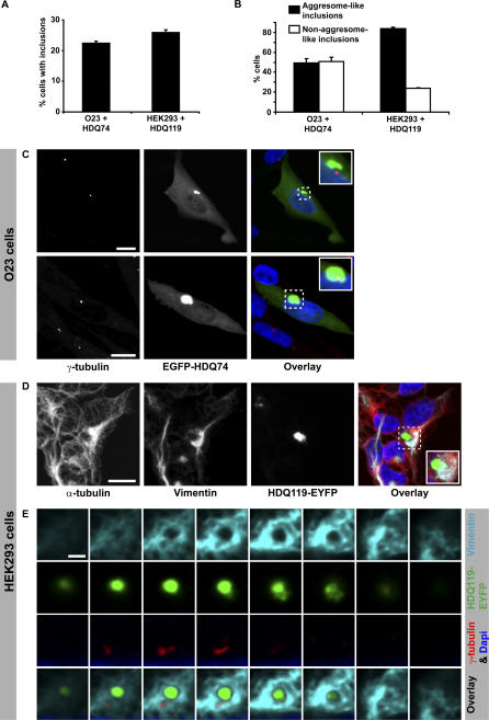
Figure 1. Polyglutamine-Expanded Proteins Form Aggresomes in Hamster O23 Cells and Human HEK293 Cells.
(A) Percentage of cells containing inclusions 24 h after transfection with a fluorescently tagged huntingtin fragment containing a stretch of either 74 (O23: EGFP-HDQ74) or 119 (HEK293: HDQ119-EYFP) glutamines.
(B) Fraction of cells showing either aggresome-like inclusions or non–aggresome-like inclusions (nuclear and/or multiple scattered inclusions). Bars represent standard errors of the mean.
(C) Aggresome-like inclusions are either close to (upper panel) or co-localise (lower panel) with the centrosomes (decorated with γ-tubulin antibodies) in interphase O23 cells (likewise in HEK293 cells, Figure S1).
(D) Vimentin microfilaments are redistributed in a cage-like manner around the inclusion, consistent with aggresome morphology. Note that also microtubules (decorated with α-tubulin antibodies) showed partial redistribution to the aggresome.
(E) Sequential confocal planes of an aggresome showing both co-localisation with the centrosome and the cage of vimentin. For a full image of this cell see Figure S1B. DNA is stained with DAPI (blue) and only shown in the overlay images. Bars in (C) and (D) represent 10 μm. Bar in (E) represents 2 μm.

