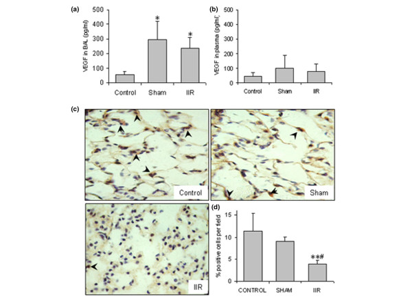
Figure 2.

Intestinal ischemia reperfusion (IIR)-induced changes in vascular endothelial growth factor (VEGF) expression in the lung. (a) VEGF in the bronchoalveolar lavage (BAL) fluid (n = 12/group); *p < 0.05 compared with the control. (b) VEGF in the plasma (n = 8/group). (c) VEGF immunostaining in the lung tissues (n = 4/group). Slides shown are representatives for each group (magnification 1,000×), and arrowheads indicate the examples of positive stained cells (in brown). (d) Quantification of VEGF positive cells per field. Ten fields were counted from each animal and four animals from each group. In the IIR group, the number and intensity of positive stained cells in the alveolar walls were remarkably decreased. **p < 0.01 compared with the control group; #p < 0.05 compared with the sham group.
