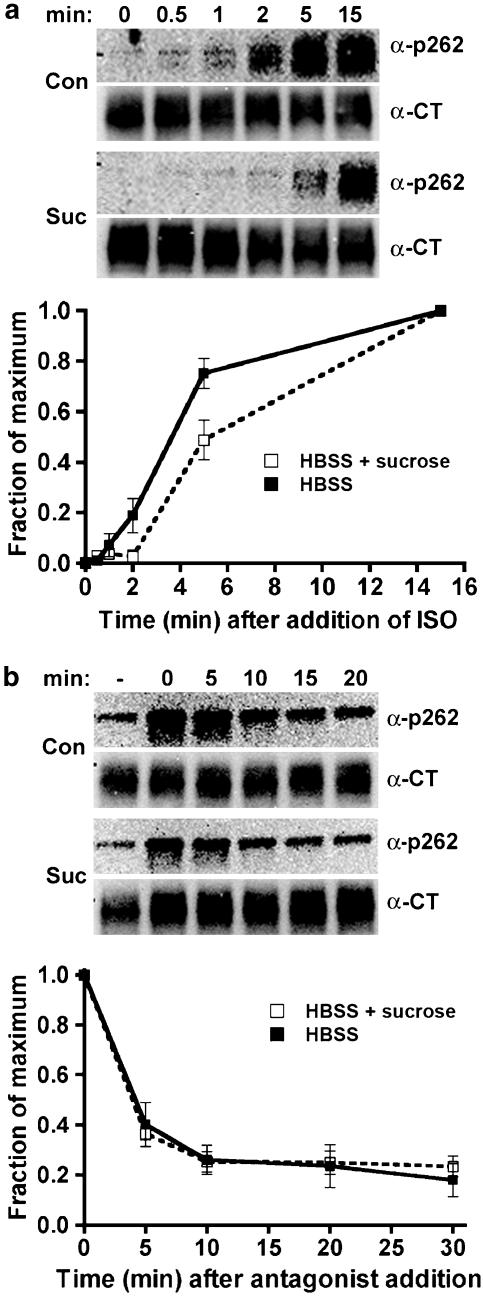
Figure 9.
Phosphorylation and dephosphorylation of β2-adrenoceptors at the PKA site Ser262 after treatment with low agonist concentration in the presence of hypertonic sucrose. 12β6 cells were grown for 24 h, then treated with HBSS alone or HBSS+0.5 M sucrose at 37°C for 30 min. (a) Phosphorylation. Isoprenaline was added to 300 pM for varying times, and then the cells were chilled and harvested in lysis buffer as described in the Methods. (b) Dephosphorylation. After 15 min of 300 pM isoprenaline treatment, the cells were washed three times with medium, then incubated for varying times in HBSS or HBSS+0.5 M sucrose containing 3 μM propranolol prior to harvest. For both (a) and (b), equivalent protein was loaded in each lane for PAGE, then immunoblotted using the antibody against phosphoserine 262 (α-p262) and visualized by chemiluminescence. After stripping the blot, it was probed with antibody to the β2-adrenoceptor C-terminus (α-CT) to normalize phosphoreceptor levels to total receptors. At the top of each panel are shown representative blots, with the data quantified as the fraction of maximum phosphorylation from three experiments shown in the lower panels (N=3).

