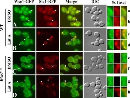
Figure 4.
Colocalization of Wsc1p but not Wsc1AAA with Sla1p. Sla1-RFP Wsc1-GFP cells (GPY4185; A and B) or Wsc1AAA-GFP (GPY4187; C and D) cells were grown to early log phase at 24°C and treated with DMSO (A and C) or 200 μM LatA (B and D) for 20 min at 24°C. Cells were analyzed by epifluorescence or DIC (4th column). Arrows indicate patches of Wsc1-GFP that colocalize with Sla1-RFP in Wsc1-GFP strains (A and B); arrowheads indicate reciprocal localization of Wsc1AAA-GFP and Sla1p-RFP in Wsc1AAA cells (C and D). Boxed areas in the merged panels are 5× magnified on the right. Insets b and g were rotated 90° counterclockwise.

