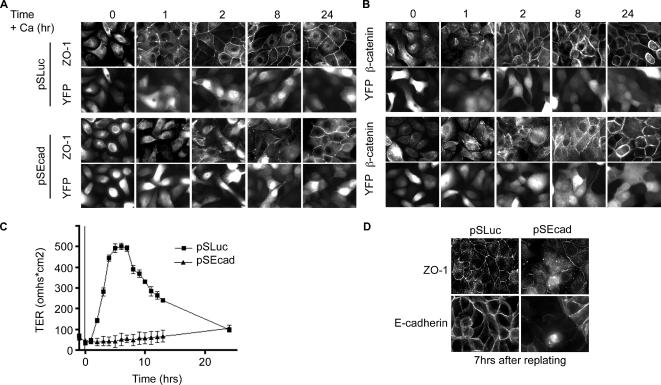
Figure 8.
TJ biogenesis is disrupted in cells depleted of E-cadherin. E-cadherin knockdown cells fail to polarize properly after calcium switch. (A) Immunofluorescence of MDCK cells 2 d after transfection with control shRNA and YFP. (B) E-cadherin–depleted cells fixed and stained 2 d after transfection. Cells were stained for ZO-1 and β-catenin. YFP marks transfected cells. (C) Average TER after calcium switch for control (■) and knockdown cells (▴). (D) Trypsinized and replated cells were analyzed by immunofluorescence for TJ formation. Cells were fixed 7 h after replating and stained for E-cadherin and ZO-1.

