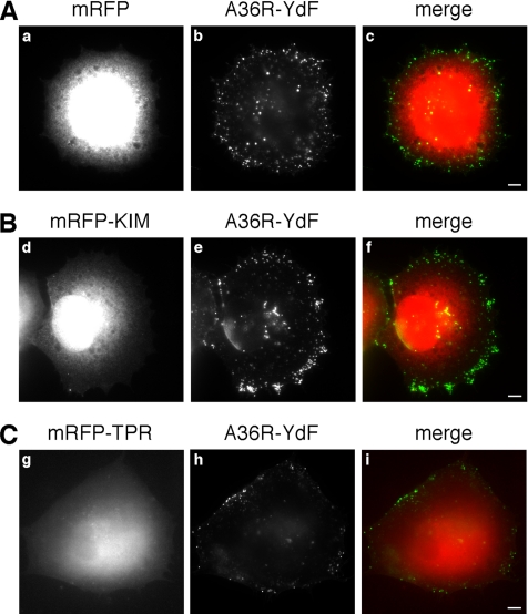
Figure 6.
The translocation of vaccinia virus to the plasma membrane is unaffected by KIM. HeLa cells were infected with the A36R-YdF vaccinia virus strain, and 4 h later they were transfected with mRFP (A), mRFP-KIM (B), or mRFP-TPR (C). After a further 4 h, cells were processed for immunofluorescence in absence of permeabilization, and extracellular virus particles were visualized. The distribution of the virus on the plasma membrane is independent of the overexpression of mRFP-KIM (compare b and e), whereas it is reduced by the overexpression of the mRFP-TPR domain (compare b and h). Bars, 5 μm.

