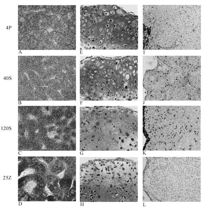
Fig. 5.

Light microscopy comparisons of 14-day chick cultures: Mineralizing controls (4P) (a, e, i), 40 nM Stauorsporine treated (b, f, j), 120 nM Staurosporine treated (c, g, k) and 25 nM Z-VAD-FMK treated (d, h, l). Alcian Blue stain (a-d; 4× original magnification): All cultures stain for proteoglycan content. The Z-VAD-fmk treated group showed strongest staining (5 d) and staurosporine treated cultures (5 b, c) showed less staining than control (5 a). Plastic epoxy sections (5 e-h; 20× magnification) stained with H&E showed distinctive differences within the hypertrophic cell population. Both treatment groups contained fewer hypertrophic cells compared to mineralizing control cultures. The Z-VAD-fmk treated group (5 h) was distinctive in that numerous cells lined the nodule surface and filled the internodular spaces. The TUNEL stain (5 i-l; 10× original magnification) showed that staurosporine contained more apoptotic cells than the mineralizing controls and higher doses of staurosporine resulted in greater apoptosis. The Z-VAD-fmk treated group (5 l) had fewer apoptotic cells than controls (5 i).
