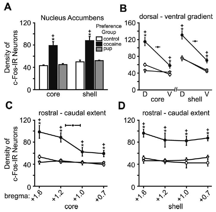
Fig. 5.

Number of c-Fos-IR neurons in the Acb core and shell subdivisions for each preference group and control. (A) Histograms show the mean (±S.E.M.) number of c-Fos-IR neurons within each subdivision. (B) Graphs show the mean (±S.E.M.) number of c-Fos-IR neurons along the dorsal-ventral gradient in the core (right) and shell (left). (C) Graphs show the mean (±S.E.M.) number of c-Fos-IR neurons along the rostral-caudal extent in the core, which were counted at four anatomic levels. (D) Graphs show the mean (±S.E.M.) number of c-Fos-IR neurons along the rostral-caudal extent in the shell, counted at the same four anatomic levels as the core. Asterisks indicate a significant difference from the control group, P<0.05. Plus signs indicate a significant difference from the other preference group, P<0.05. Statistical details in the Results.
