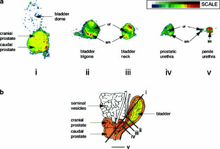
Figure 1.
Presence of α1A-adrenoceptor protein in the lower urinary tract of the monkey. Receptors were localized by autoradiography, using [3H]praozsin and defining non-specific binding in the presence of SNAP 5272. Receptor autoradiograms were scanned into computer as a 16 grey scale image. The 16 grey levels corresponding to specific α1A-adrenoceptor receptor binding were each assigned colour (see scale) to allow subtle differences in film exposure to be easily visible. Sections show bladder dome and prostate (i), bladder trigone (ii), bladder base (iii), prostatic urethra (iv) and penile urethra (v). Key: sm (smooth muscle); ur (urothelium); lm (longitudinal muscle). Schematic representation of monkey urinary tract together with the orientation of sectioning planes (i)–(v) shown in (b). Taken with permission from Walden et al. (1997).

