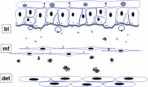
Figure 5.
A schematic diagram showing the main ultrastructural constituent elements of the superficial layers of the human bladder. Flattened cells with pale nuclei, known as ‘umbrella cells' form the innermost layer of the bladder with tight and adhaerens junctions linking them to one another. These are supported by basal epithelial cells which are closely attached to the basal lamina (bl). Naked axons can be seen between the basal processes of the epithelial cells and immediately beneath the basal lamina. In the zone immediately beneath the epithelial basal lamina are fine axons, either naked or in intimate association with flattened cells have the cytological characteristics of myofibroblasts (mf). Also in this layer are fenestrated capillaries orientated towards the urothelial surface. Within the deeper zones of the lamina propria is a diffuse plexus of unmyelinated fibres containing slender axons linking periodic varicosities which enclose numerous small clear and dense cored vesicles. Close to the smooth muscle of the detrusor (det) nerves consist of small myelinated fibres and numerous unmyelinated strands, partially or completed invested by perineurium. From (Fowler et al., 2002) with permission.

