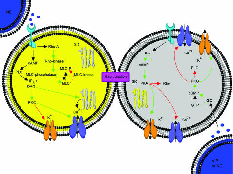
Figure 1.
Schematic depiction of the physiological/pharmacological basis for contraction and relaxation of corporal myocytes. Mechanisms are summarized from the text and cited literature throughout this report. Where SR: sarcoplasmic reticulum, PLC: phospholiapse C, DAG: diacylglycerol, PKC: protein kinase C, IP3: inositol trisphosphate, MLC: myosin light chain. Stimulatory pathways are illustrated via the green arrows, while the red arrows represent inhibitory regulation.

