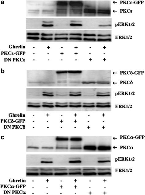
Figure 8.
PKCɛ but not PKCδ is involved in hGHSR-1a-mediated activation of ERK1/2. (a) CHO cells were transiently transfected with plasmid encoding hGHSR-1a (10 μg) together with 20 μg of empty pEGFP-N1 vector, wild-type PKCɛ-GFP or c-myc-tagged DN PKCɛ. Cells, which had been serum-starved for 1 h were stimulated with ghrelin (100 nM, 5 min). (b) CHO cells were transiently transfected with plasmid DNA encoding hGHSR-1a (10 μg) together with 20 μg of empty pEGFP-N1 vector, wild-type PKCδ-GFP or c-myc-tagged DN PKCδ. Cells that had been serum-starved for 1 h were stimulated with ghrelin (100 nM, 5 min). (c) CHO cells were transiently transfected with plasmid DNA encoding hGHSR-1a (10 μg) together with 20 μg of empty pEGFP-N1 vector, wild-type PKCα-GFP or c-myc-tagged DN PKCα. Cells, which had been serum-starved for 1 h, were then stimulated with ghrelin (100 nM, 5 min). Phosphorylated ERK1/2 (pERK1/2) and total ERK1/2 (ERK1/2) were detected by immunoblotting as described in Figure 2. The expression level of the different PKC isoforms was revealed by probing the western blot with anti-PKCα, δ or ɛ antibodies. All blots are representative of three experiments with similar results.

