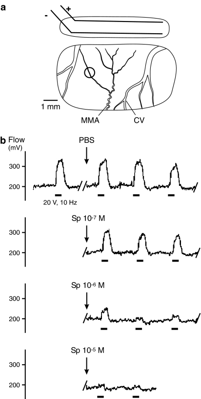
Figure 1.
(a) Stimulation and recording window in the parietal bone showing bipolar electrodes (±) and the recording site (ring) over a branch of the MMA; cortical veins (CV). (b) Sections from a continuous recording of meningeal blood flow showing increases in flow upon electrical stimulation (bars) and their changes after topical application of PBS and Spiegelmer NOX-C89 (10−7–10−5 M).

