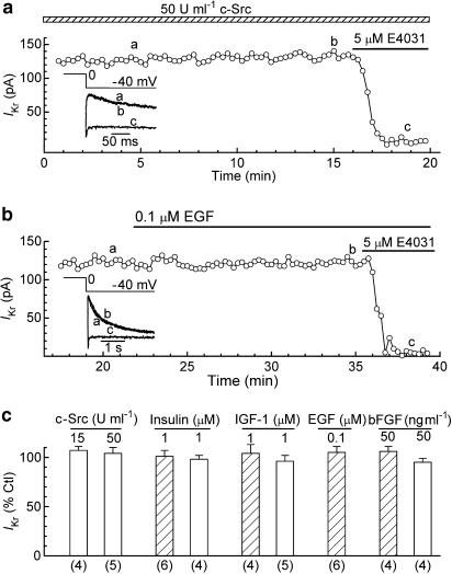
Figure 6.
Effects of stimulators of tyrosine phosphorylation on IKr in ruptured-patch and perforated-patch myocytes. The myocytes were pulsed from −40 to 0 mV for 200 ms for measurement of the amplitude of the IKr tail on repolarisation to −40 mV before (control) and 10–15 min after the initiation of treatment. (a) IKr in a ruptured-patch myocyte dialysed with pipette solution that contained 50 U ml−1 c-Src. Inset: superimposed records obtained at the times indicated in the plot. (b) IKr in a perforated-patch myocyte treated with 0.1 μM EGF. Inset: superimposed records obtained at the post-pipette-attachment times indicated in the plot. (c) Summary of the data obtained on ruptured-patch (open bars) and perforated-patch (hatched bars) myocytes. In the experiments with c-Src dialysate, the amplitude of IKr measured during the first minute of dialysis was taken as the control amplitude. Numbers of myocytes in parentheses.

