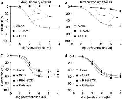
Figure 2.
Relaxant effect of acetylcholine in extrapulmonary (a, c) and intrapulmonary (b, d) arteries from control mice (wild-type). The effect of acetylcholine was studied in the absence or presence of 300 μM L-NAME, 1 μM ODQ (a, b), 200 U ml−1 SOD, 120 U ml−1 PEG-SOD or 250 U ml−1 catalase (c, d). In each panel, the response is expressed as the percentage of relaxation of the tone induced by PGF2α. *P<0.05, ***P<0.001: significant difference compared to controls (ANOVA). n=3–15 (a, b) or 5–15 (c, d).

