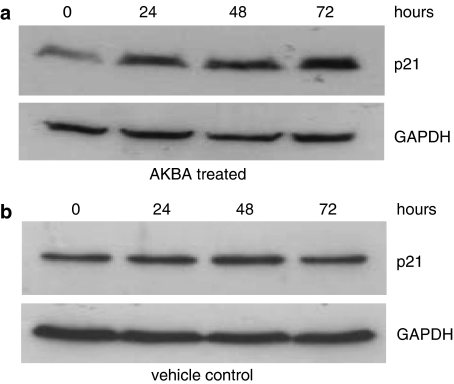
Figure 6.
Expression of p21 in AKBA treated cells. HCT-116 cells were treated with 20 μM AKBA for 0, 24, 48 and 72 h. The expression of p21 was assayed by Western blot analysis. GAPDH was used as a housekeeping control. (a) Expression of p21 in HCT-116 cells after treatment with AKBA; (b) expression of p21 in HCT-116 cells treated with vehicle control. Results shown are representative of at least three separate experiments.

