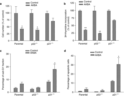
Figure 8.
Effect of AKBA on growth and apoptosis in parental, p21−/− and p53−/− HCT-116 cells. Cells were treated with 20 μM AKBA for 48 h. Control cells were treated with 0.1% ethanol. The number of cells was counted using a hemacytometer. DNA synthesis was assayed by 3H-thymidine incorporation. Apoptosis was assayed by both cell cycle analysis and annexin V staining. (a) Changes in cell numbers after treatment with AKBA in parental, p21−/− and p53−/− HCT-116 cells; (b) changes in DNA synthesis in parental, p21−/− and p53−/− HCT-116 cells after treatment with AKBA; (c) changes in sub-G1 fraction after treatment with AKBA in parental, p21−/− and p53−/− HCT-116 cells; (d) changes in apoptotic rate assayed by annexin V and propidium iodide double staining in parental, p21−/− and p53−/− HCT-116 cells. Results shown are representative of three separate experiments. *P<0.05 compared to control. **P<0.01 compared to control.

