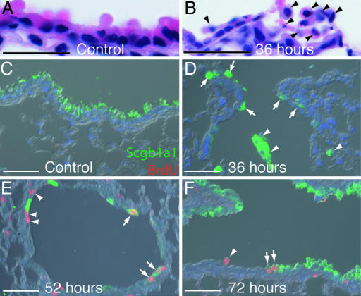
Fig. 3.
Characterization of the extent of the naphthalene injury and timing of proliferation. (A–F) Paraffin sections of bronchiole epithelium from male mice. (A and B) Hematoxylin and eosin staining. (A) Thirty-six hours after control corn oil injection. (B) Thirty-six hours after naphthalene. Arrowheads mark cells with no attachment to the basal lamina. (C–F) Anti-Scgb1a1 (green), anti-BrdU (red), and DAPI (blue). (C) Fifty-two hours after control injection. Clara cells predominate the airway epithelium and no dividing cells are present. (D) Thirty-six hours after naphthalene. Clara cells have detached from the basal lamina (arrowheads), but small numbers are retained at the BADJ (arrows) and at intervals throughout the bronchioles (data not shown). Cells have not yet started to divide. (E) Fifty-two hours after naphthalene injection. Dividing epithelial cells are observed; they are either Scgb1a1+ (arrows) or Scgb1a1− (arrowheads). (F) Seventy-two hours after naphthalene injection. Dividing Scgb1a1+ (arrows) and Scgb1a1− (arrowhead) cells are still observed, and many more Clara cells are also present. (Scale bars: A and B, 20 μm; C–F, 200 μm.)

