
Figure 3.
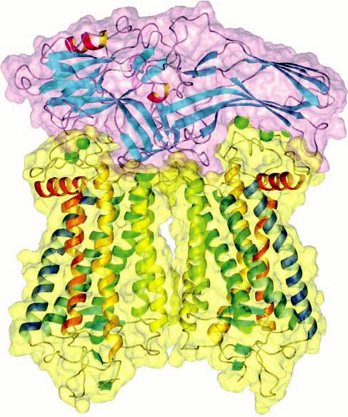
Ribbon and space-filling model of a complex between a rhodopsin dimer and a monomer of arrestin. Arrestin (purple) has a bipartite structure that can accommodate two molecules of rhodopsin (yellow). Phosphorylation sites on rhodopsin are represented by green spheres (adapted from ref 100).