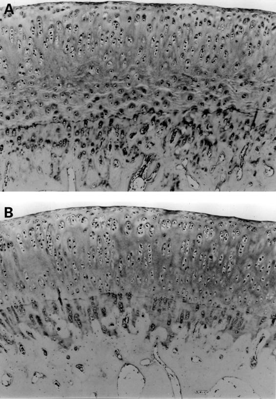
Figure 6 .

(A) At 32 weeks, cartilage is oedematous and many splits and wrinkles of matrix are evident in intermediate zones of patellar. Chondrocytes of superficial zone are not morphologically different to chondrocytes of control. (Safranin-O, original magnification × 100). (B) Patellar cartilage at 32 weeks in control. No remarkable changes noted in chondrocytes and matrix. (Safranin-O, original magnification × 100).
