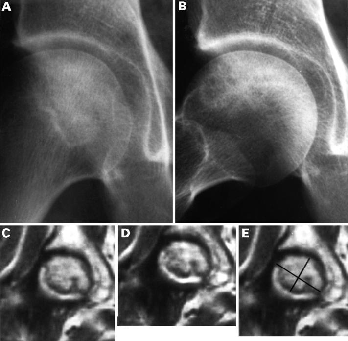
Figure 1 .
Case 2 , 56 year old women. Right hip: (A) on AP view a linear sclerotic line at the junction of femoral head and neck delineates a sclerotic area in the centre of the head; (B) on profile, the sclerotic zone is limited to the anterosuperior part of the head. On both views the femoral head is spherical (stage II); (C) MRI, T1 weighted image (TE: 500 ms, TR: 34 ms) a concave superiorly low signal intensity line delineates the necrotic area; (D) T2 weighted image (TE: 2000 ms, TR: 50 ms) the same line is observed, with a high signal intensity band at the convex aspect of the line ("double rim sign"); (E) on the T1 weighted image, the necrotic area is assimilated to a rectangle whose length and width are measured with a magnifying glass graduated in tenths of a millimetre.

