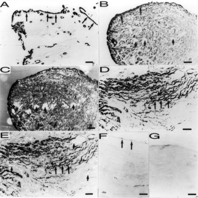
Figure 1 .

Immunoreactivity for tenascin-C in the synovial membrane-like interface tissue from aseptic loosening of THR and control synovial samples (APAAP staining, original magnification × 250). (A) Immunoreactivity for tenascin-C in the synovial membrane-like interface tissue. Tenascin-C staining was very strong in the synovial lining-like layer (arrows) of interface tissue. This sample/ section also contains intervening matrix, which does not stain for tenascin. (B)Tenascin-C was detected in all fields/extracellular matrix of the synovial membrane-like interface tissue in this revision THR patient. Particularly strong staining was found in the synovial lining-like layer (larger arrows). Heavy deposits of debris (see (C) for verification) were found embedded in the tissue (small arrows). (C) Same section as in (B) photographed with polarised light shows that there were many birefringent polyethylene particles embedded in the tissue (some are marked with arrows). (D) Fibroblasts and their pericellular matrix (arrows) showed usually intense tenascin expression in the synovial membrane-like interface tissue samples, whereas the extracellular collagenous matrix did not stain as strongly (the white intervening areas). Note, that fibroblasts send long and slender extensions, which pass between the collagenous fibres of the connective tissue and would be difficult to identify without tenascin-C staining. (E) Same section as in (D) photographed with polarised light shows polyethylene deposits (some are marked with small arrows). (F) Very weak tenascin-C staining in the control synovial sample from osteoarthritis. (G) For staining control the specific primary IgG2a antibody were replaced with monoclonal IgG with an irrelevant specificity (Aspergillus niger glucose oxidase), but of the same subtype and concentration as the specific primary antibody. Comparison with the (A) confirms the specificity of the staining.
