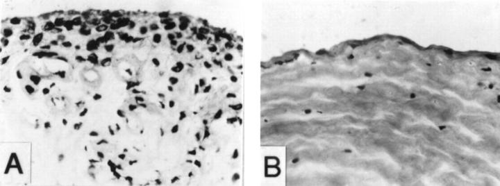
Figure 2 .
Histological examination of the interface tissue samples with different staining scores (haematoxylin and eosin staining, original magnification × 250). (A) Macrophage-like cells accumulation in a synovial membrane-like interface tissue sample with high staining score (staining score = 4). (B) Matrix fibrosis in the interface tissue sample with lower staining score (staining score = 3).

