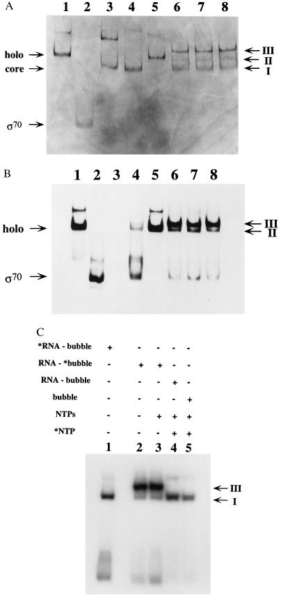
Figure 4.
Binding of RNA primer and σ70 to synthetic transcription complexes is mutually exclusive. (A) Holo RNA polymerase (1.2 pmols), 1.4 pmols of core RNA polymerase, and 2 pmols of purified sigma subunit (provided by Lam Nguyen) were resolved on a nondenaturing gel (Materials and Methods) in the absence and presence of nucleic acids and were visualized by silver staining of the protein components. Lane 1 contains holo RNA polymerase; lane 2 contains sigma subunit; lane 3 contains core RNA polymerase; lane 4 contains holo RNA polymerase plus 2 μg of yeast tRNA; lane 5 contains holo RNA polymerase plus 35 pmols of RNA primer that is 12 nt in length; lane 6 contains holo RNA polymerase plus 1.4 pmols RNA–DNA bubble duplex; lane 7 contains holo RNA polymerase plus RNA–DNA bubble duplexes and 100 μM concentrations of ATP, CTP, and UTP; lane 8 contains holo RNA polymerase plus 1.7 pmols of DNA bubble duplex lacking the RNA primer. The gel positions of the monomeric forms of holo, core, and sigma subunit are marked on the left side of the gel, whereas the positions of gel bands I, II, and III are marked on the right. (B) Staining of a gel identical to that shown in Fig. 4A with a sigma subunit monoclonal antibody (see Materials and Methods). (C) An autoradiogram of a nondenaturing gel, similar to those shown in Fig. 4 A and B, in which 1.2 pmols of holo RNA polymerase were incubated with radiolabeled RNA–DNA bubble duplexes in 10-μl reaction volumes. Lane 1 contains 0.7 pmols of 5′-end labeled RNA primer plus unlabeled DNA bubble duplex; lane 2 contains 0.5 pmols of a DNA bubble duplex radiolabeled on the bottom strand plus a nonlabeled RNA primer; lane 3 contains the same materials as lane 2 plus 100 μM of ATP, CTP, and UTP; lane 4 contains 0.45 pmols of a nonlabeled RNA–DNA bubble duplex in the presence of 100 μM ATP, 100 μM UTP, and 1.5 μM 32P-α CTP; lane 5 contains 0.55 pmols of a nonlabeled DNA bubble duplex (lacking an RNA primer) plus 100 μM ATP, 100 μM UTP, and 1.5 μM 32P-αCTP. The radiolabeled species are marked (∗) at the top of the lanes, and the positions of bands I and III are marked on the right side of the gel.

