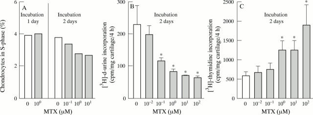
Figure 1 .
Effects of MTX on the DNA metabolism of cultured articular cartilage. (A) Reduction of the proportion of chondrocytes in S-phase by MTX in vitro. Bovine articular cartilage explants were cultured in the presence of various MTX concentrations, then isolated with collagenase, stained with propidium iodide, and analysed for DNA content by flow cytometry. The proportion of chondrocytes in S-phase was dose dependently reduced by MTX after 48 hours of culture (p<0.05; linear regression analysis), but not after 24 hours. Bars represent means of at least 10 000 events. (B) Inhibition of [3H]-d-uridine-incorporation into chondrocytes by MTX in vitro. Bovine articular cartilage explants were cultured in the presence of various MTX concentrations for two days, then labelled with [3H]-d-uridine for four hours, washed in PBS to remove free [3H]-d-uridine, digested with papain, and counted for [3H]. Bars represent means and SEM of eight cartilage samples per group; * p < 0.05 v control without MTX (t test). (C) Stimulation of [3H]-thymidine incorporation into chondrocytes by MTX in vitro. Bovine articular cartilage explants were cultured in the presence of various MTX concentrations for two days, then labelled with [3H]-thymidine for four hours, washed in PBS to remove free [3H]-thymidine, digested with papain, and counted for [3H]. Bars represent means and SEM of eight cartilage samples per group; * p<0.05 v control without MTX (t test).

