
Figure 2 .
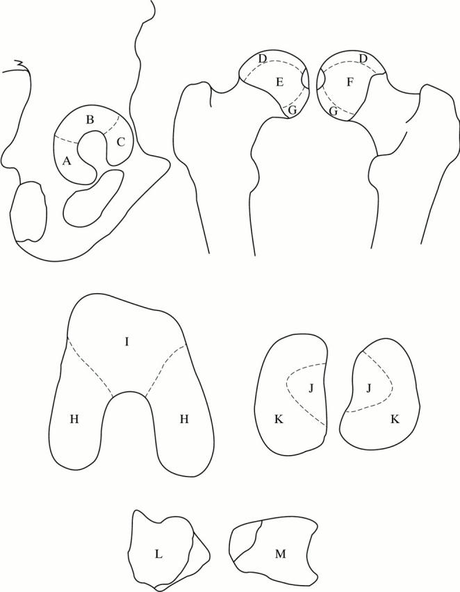
Distinct areas of the lower limb joints (A, B, C: posterior, superior, and anterior areas of the acetabulum respectively; D, E, F, G: superior, anterior, and posterior areas of the femoral head respectively; H, I: patellar surface and femoral condyles respectively; J, K: tibial areas of the knee covered by the menisci and those that come into direct contact with the femur respectively; L, M: talar and tibial areas of the ankle respectively).