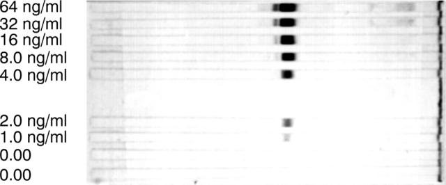
Figure 2 .
Illustration of the use of chimeric IgA anti-NIP antibodies to semiquantify immunoblotting data. BSA-NIP conjugate was separated on SDS-PAGE and transferred to a blotting membrane. After incubation with different concentrations of IgA anti-NIP antibodies (0-64 ng/ml) the membrane was incubated with anti-IgA enzyme conjugate and developed with BCIP/NBT for 30 minutes.

