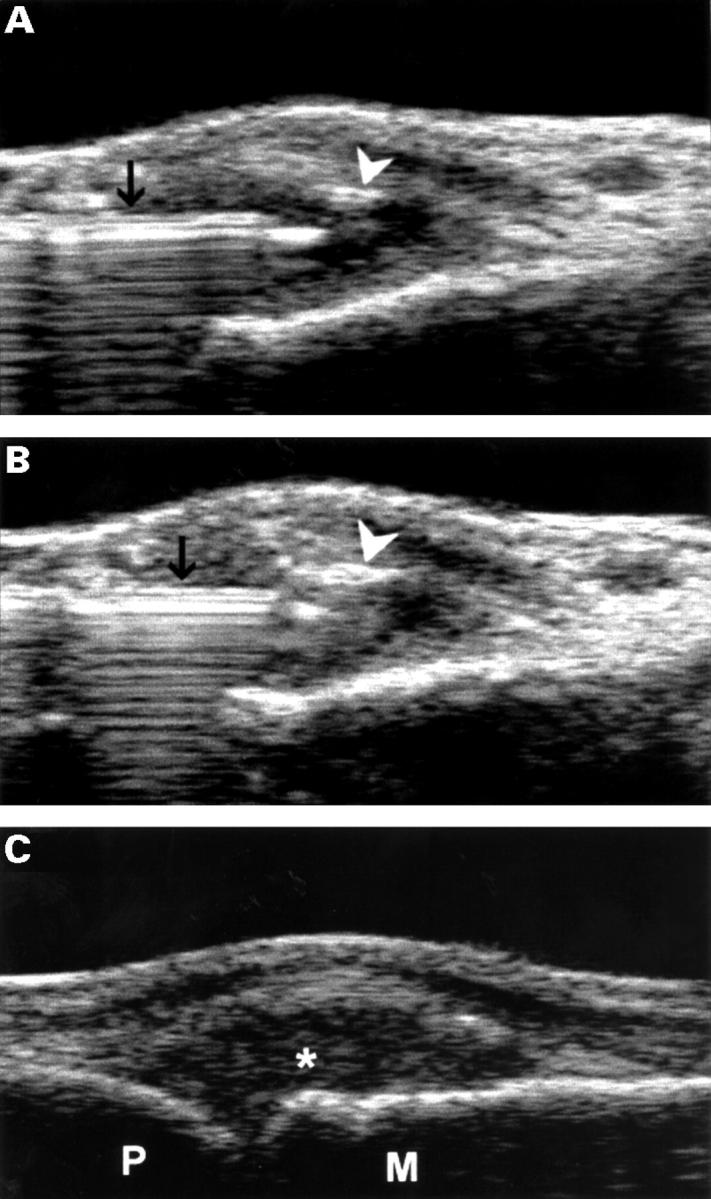
Figure 5 .

(A) Longitudinal sonogram of the MCP joint showing the placement of the tip of the needle within the joint cavity. The image has been obtained after the injection with a minimal amount of triamcinolone hexacetonide. Triamcinolone drops are highly echoic and can be easily visualised as white spots (arrowhead). Black arrow = needle. (B) Complete filling of the joint cavity by corticosteroid. (C) Longitudinal sonogram of the MCP joint after the injection shows an echoic joint cavity (*). M = metacarpal bone; P = proximal phalanx.
