Abstract
OBJECTIVES—It has been suggested that the humoral immune system plays a part in the pathogenesis of pulmonary fibrosis. Although circulating autoantibodies to lung protein(s) have been suggested, few lung proteins have been characterised. The purpose of this study is to determine the antigen recognised by serum of a patient with pulmonary fibrosis associated with dermatomyositis. METHODS—To accomplish this, anti-small airway epithelial cell (SAEC) antibody in a patient's serum was evaluated using a western immunoblot. RESULTS—An autoantibody against SAEC was found, and the antigen had a molecular weight of 62 kDa. Using the patient's serum, clones from the normal lung cDNA library were screened and demonstrated that anti-SAEC antibody in the patient's serum was against ADAM (A disintegrin and metalloprotease) 10. CONCLUSION—This is the first report that demonstrates the existence of anti-ADAM 10 antibody in a patient with pulmonary fibrosis associated with dermatomyositis.
Full Text
The Full Text of this article is available as a PDF (771.2 KB).
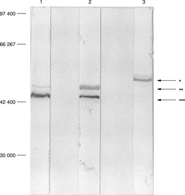
Figure 1 .
Western immunoblot analysis using the patient's sera against lysates of PC9, A549, and SAEC cell lines. Lane 1; PC9, lane 2; A549; lane 3; SAEC cell line. In SAEC cell line, only one band (arrow *) that has a molecular weight of 62 kDa, which is the same as ADAM 10, is demonstrated. In PC9 and A549 cell lines, two bands (arrows ** and ***) are demonstrated. One antigen, which has a molecular weight of 54 kDa (arrow **), was previously identified as cytokeratin 8. Arrow *** is not characterised.
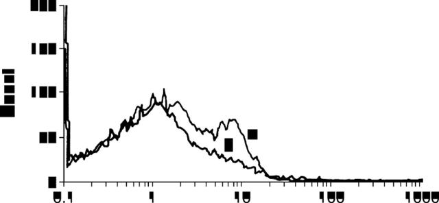
Figure 2 .
Immunofluorescence staining of ADAM 10 expressing COS-7 cells by a patient's serum. This histogram demonstrates that COS-7 cells transfected with the full length of ADAM 10 are stained positively by FY serum (a). In contrast, COS-7 cells transfected with pCDNA1.1/Amp alone were unstained with FY serum (b).


