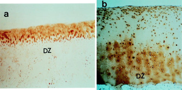
Figure 1 .
Immunohistochemical identification of type II collagen degradation in rheumatoid articular cartilage. Tibial plateau articular cartilage (woman 52 years) taken apart from joint margins were fixed for six hours at 4°C in 2% paraformaldehyde containing 0.075 M lysine and 0.01 M sodium periodate solution, and washed at 4°C with 0.01 M phosphate buffer saline (PBS, pH 7.2) containing glycerol, as previously described by McLean and Nakano.19 Then they were decalcified with EDTA-glycerol solution at −5°C. The samples were embedded in paraffin wax, and immunohistochemical analysis was assessed on the sections using the avidin-biotin-peroxidase complex (ABC) immunoperoxidase.20 The cartilage sections were stained with monoclonal antibody against human type II collagen (a) and rat polyclonal antibody against CNBr derived peptides of type II collagen (b). Less staining for type II collagen and intense staining for CNBr derived peptides of type II collagen in the deep zone (DZ) matrix are observed. ((a) Original magnification × 6.6, (b) original magnification × 13.2).

