Abstract
OBJECTIVES—To determine the difference in shape of the distal femur, viewed axially in two dimensions, between eburnated and non-eburnated femora. METHODS—A comparison of 52 non-eburnated and 16 eburnated femora drawn from a large archeological skeletal population. Eburnation was taken to indicate late stage osteoarthritis. Shape variability, based on landmarks, was quantified using a principal components analysis after a Procrustes alignment. RESULTS—A statistically significant difference was found between the two groups. This was with respect to the patellar groove and the shape of the medial condyle. The latter difference is consistent with bone remodelling as a knee stabilising mechanism. CONCLUSIONS—Anatomical shape can be quantified using an uncomplicated statistical technique. It was used to quantify the shape of the distal femur and demonstrate shape differences associated with osteoarthritis of the knee. Keywords: osteoarthritis; knee; bone remodelling
Full Text
The Full Text of this article is available as a PDF (160.1 KB).
Figure 1 .
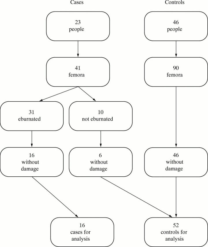
Illustration of selection of study material.
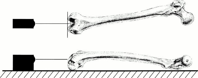
Figure 2 .
Alignment of femora to camera: femora are allowed to rest naturally upon the trochanters and most posterior points of the condyles.
Figure 3 .
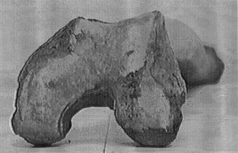
Example of bitmap digitised from video image.
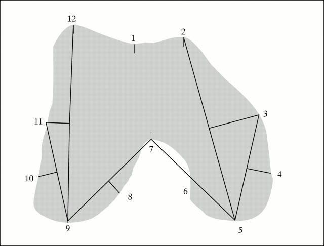
Figure 4 .
Location of 12 landmarks used for analysis. Landmarks 1, 2, 5, 7, 9, and 12 were located by hand. The remaining six landmarks were defined relative to these.
Figure 5 .
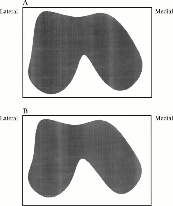
Mean configurations. Periodic splines were used to produce outlines for the 12 landmark mean configurations. The mean shape of the 16 eburnated femora is shown (A) compared with that of the 52 non-eburnated (B).
Figure 6 .
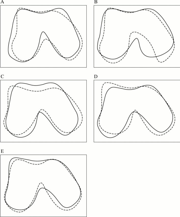
Effect of principal components on the overall mean. The mean configuration plus (solid line) or minus (dotted line) three standard deviations of the first five principal components (A)-(E). A significant difference was found between the two groups with respect to the second principal component scores. The eburnated femora tended to have higher second principal component scores.
Figure 7 .
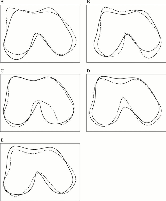
Effect of varimax rotated principal components on the overall mean. The mean configuration plus (solid line) or minus (dotted line) three standard deviations of the rotated first five principal components (A)-(E). A significant difference was found between the two groups with respect to the second and third principal component scores. The eburnated femora tended to have higher scores for both of these rotated principal components.
Selected References
These references are in PubMed. This may not be the complete list of references from this article.
- Brage M. E., Draganich L. F., Pottenger L. A., Curran J. J. Knee laxity in symptomatic osteoarthritis. Clin Orthop Relat Res. 1994 Jul;(304):184–189. [PubMed] [Google Scholar]
- Bullough P. G. Osteoarthritis: pathogenesis and aetiology. Br J Rheumatol. 1984 Aug;23(3):166–169. doi: 10.1093/rheumatology/23.3.166. [DOI] [PubMed] [Google Scholar]
- Bullough P. G. The geometry of diarthrodial joints, its physiologic maintenance, and the possible significance of age-related changes in geometry-to-load distribution and the development of osteoarthritis. Clin Orthop Relat Res. 1981 May;(156):61–66. [PubMed] [Google Scholar]
- Casscells S. W. Gross pathological changes in the knee joint of the aged individual: a study of 300 cases. Clin Orthop Relat Res. 1978 May;(132):225–232. [PubMed] [Google Scholar]
- Cooke D., Scudamore A., Li J., Wyss U., Bryant T., Costigan P. Axial lower-limb alignment: comparison of knee geometry in normal volunteers and osteoarthritis patients. Osteoarthritis Cartilage. 1997 Jan;5(1):39–47. doi: 10.1016/s1063-4584(97)80030-1. [DOI] [PubMed] [Google Scholar]
- Cooper C., McAlindon T., Snow S., Vines K., Young P., Kirwan J., Dieppe P. Mechanical and constitutional risk factors for symptomatic knee osteoarthritis: differences between medial tibiofemoral and patellofemoral disease. J Rheumatol. 1994 Feb;21(2):307–313. [PubMed] [Google Scholar]
- Dieppe P., Kirwan J. The localization of osteoarthritis. Br J Rheumatol. 1994 Mar;33(3):201–203. doi: 10.1093/rheumatology/33.3.201. [DOI] [PubMed] [Google Scholar]
- Felson D. T., Anderson J. J., Naimark A., Walker A. M., Meenan R. F. Obesity and knee osteoarthritis. The Framingham Study. Ann Intern Med. 1988 Jul 1;109(1):18–24. doi: 10.7326/0003-4819-109-1-18. [DOI] [PubMed] [Google Scholar]
- Felson D. T., Naimark A., Anderson J., Kazis L., Castelli W., Meenan R. F. The prevalence of knee osteoarthritis in the elderly. The Framingham Osteoarthritis Study. Arthritis Rheum. 1987 Aug;30(8):914–918. doi: 10.1002/art.1780300811. [DOI] [PubMed] [Google Scholar]
- Felson D. T., Radin E. L. What causes knee osteoarthrosis: are different compartments susceptible to different risk factors? J Rheumatol. 1994 Feb;21(2):181–183. [PubMed] [Google Scholar]
- Good L., Odensten M., Gillquist J. Intercondylar notch measurements with special reference to anterior cruciate ligament surgery. Clin Orthop Relat Res. 1991 Feb;(263):185–189. [PubMed] [Google Scholar]
- Harrison M. M., Cooke T. D., Fisher S. B., Griffin M. P. Patterns of knee arthrosis and patellar subluxation. Clin Orthop Relat Res. 1994 Dec;(309):56–63. [PubMed] [Google Scholar]
- Hart D. J., Spector T. D. The relationship of obesity, fat distribution and osteoarthritis in women in the general population: the Chingford Study. J Rheumatol. 1993 Feb;20(2):331–335. [PubMed] [Google Scholar]
- Jacobsen K. Osteoarthrosis following insufficiency of the cruciate ligaments in man. A clinical study. Acta Orthop Scand. 1977;48(5):520–526. doi: 10.3109/17453677708989742. [DOI] [PubMed] [Google Scholar]
- Jurmain R. D., Kilgore L. Skeletal evidence of osteoarthritis: a palaeopathological perspective. Ann Rheum Dis. 1995 Jun;54(6):443–450. doi: 10.1136/ard.54.6.443. [DOI] [PMC free article] [PubMed] [Google Scholar]
- KELLGREN J. H., LAWRENCE J. S. Radiological assessment of osteo-arthrosis. Ann Rheum Dis. 1957 Dec;16(4):494–502. doi: 10.1136/ard.16.4.494. [DOI] [PMC free article] [PubMed] [Google Scholar]
- Lane N. E. Physical activity at leisure and risk of osteoarthritis. Ann Rheum Dis. 1996 Sep;55(9):682–684. doi: 10.1136/ard.55.9.682. [DOI] [PMC free article] [PubMed] [Google Scholar]
- Lovász G., Llinás A., Benya P., Bodey B., McKellop H. A., Luck J. V., Jr, Sarmiento A. Effects of valgus tibial angulation on cartilage degeneration in the rabbit knee. J Orthop Res. 1995 Nov;13(6):846–853. doi: 10.1002/jor.1100130607. [DOI] [PubMed] [Google Scholar]
- Marshall J. L., Olsson S. E. Instability of the knee. A long-term experimental study in dogs. J Bone Joint Surg Am. 1971 Dec;53(8):1561–1570. [PubMed] [Google Scholar]
- Moussa M. Rotational malalignment and femoral torsion in osteoarthritic knees with patellofemoral joint involvement. A CT scan study. Clin Orthop Relat Res. 1994 Jul;(304):176–183. [PubMed] [Google Scholar]
- Pottenger L. A., Phillips F. M., Draganich L. F. The effect of marginal osteophytes on reduction of varus-valgus instability in osteoarthritic knees. Arthritis Rheum. 1990 Jun;33(6):853–858. doi: 10.1002/art.1780330612. [DOI] [PubMed] [Google Scholar]
- Rogers J., Shepstone L., Dieppe P. Bone formers: osteophyte and enthesophyte formation are positively associated. Ann Rheum Dis. 1997 Feb;56(2):85–90. doi: 10.1136/ard.56.2.85. [DOI] [PMC free article] [PubMed] [Google Scholar]
- Spector T. D., Hart D. J., Doyle D. V. Incidence and progression of osteoarthritis in women with unilateral knee disease in the general population: the effect of obesity. Ann Rheum Dis. 1994 Sep;53(9):565–568. doi: 10.1136/ard.53.9.565. [DOI] [PMC free article] [PubMed] [Google Scholar]
- Wu D. D., Burr D. B., Boyd R. D., Radin E. L. Bone and cartilage changes following experimental varus or valgus tibial angulation. J Orthop Res. 1990 Jul;8(4):572–585. doi: 10.1002/jor.1100080414. [DOI] [PubMed] [Google Scholar]
- Yoshioka Y., Siu D., Cooke T. D. The anatomy and functional axes of the femur. J Bone Joint Surg Am. 1987 Jul;69(6):873–880. [PubMed] [Google Scholar]
- van Saase J. L., van Romunde L. K., Cats A., Vandenbroucke J. P., Valkenburg H. A. Epidemiology of osteoarthritis: Zoetermeer survey. Comparison of radiological osteoarthritis in a Dutch population with that in 10 other populations. Ann Rheum Dis. 1989 Apr;48(4):271–280. doi: 10.1136/ard.48.4.271. [DOI] [PMC free article] [PubMed] [Google Scholar]


