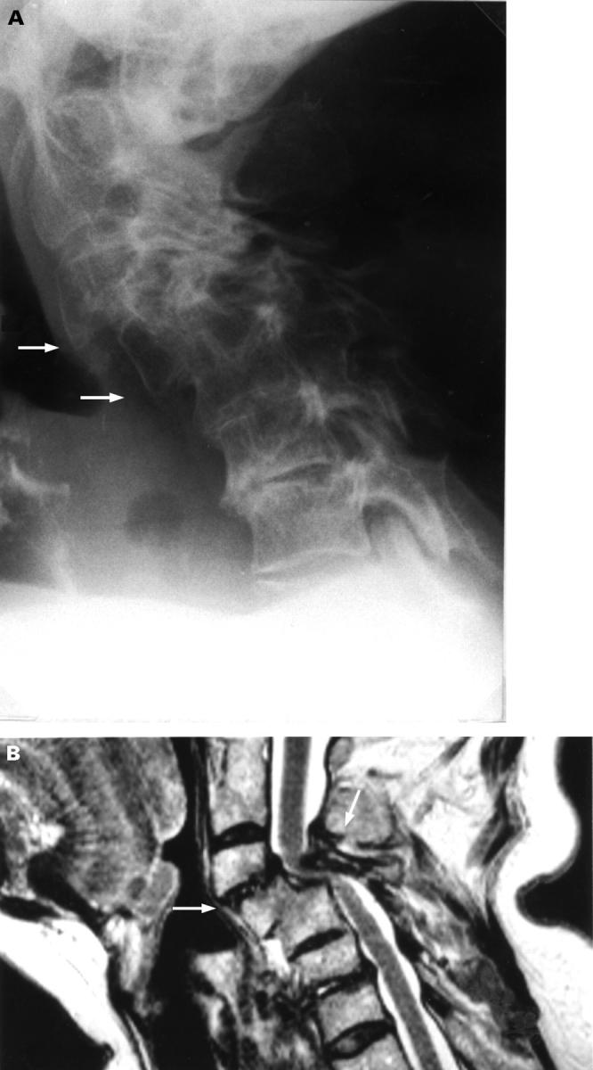
Figure 3 .

Lateral plain radiography in flexion (A) and T2 weighted sagittal MR image (B) of the cervical spine showing C3-C4 subluxation of 9 mm, stable C4-C5 subluxation of 8 mm with fusion of the vertebral bodies of C4 and C5, narrowing of the spinal subarachnoid space, and compression of the spinal cord.
