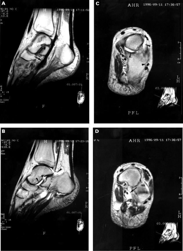
Figure 2 .
MRI of the foot of RS3PE syndrome. (A) Sagittal T1 weighted section shows marked tenosynovitis of tibialis anterior tendon (black arrow). (B) Sagittal T2 weighted section shows marked tenosynovitis of tibialis anterior tendon (black arrow), ankle joint effusion extending into Kager's triangle (black/white arrow) and talonavicular joint effusion (black/white arrow). (C) Coronal T2 weighted section at the apex of the talus shows talonavicular joint effusion (black/white arrow), tenosynovitis of flexor hallucis longus tendon (double open arrowhead), tenosynovitis of flexor digitorum longus tendon (open arrowhead), tenosynovitis of tibialis posterior tendon (open white arrow) and tenosynovitis of peroneal tendons (black arrowhead). (D) Coronal T2 weighted section through the navicular bone shows marked tenosynovitis of tibialis anterior tendon (black arrow).

