
Figure 1 .
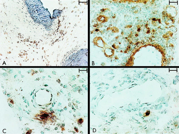
Videoprint photographs illustrating brown (diaminobenzidine) immunoperoxidase staining for cytokine producing cells in cryopreserved synovial membrane biopsy specimens obtained from patients with rheumatoid arthritis (RA). The cells were counterstained with haematoxylin. (A) Interleukin 1β producing cells in pannus tissue penetrating bone (dark blue staining) (original magnification ×200, bar represents 40 µm). (B) Interleukin 1α production occurring in both vascular endothelial cells and individual macrophages (original magnification ×320, bar represents 32 µm). (C) Tumour necrosis factor α (TNFα) producing cells in the sublining layer with additional extracellular TNFα staining encompassing producer cells (original magnification ×400, bar represents 20 µm). (D) Interleukin 6 production was seen in scattered cells with minimum additional extracellular staining (original magnification ×400, bar represents 20 µm).