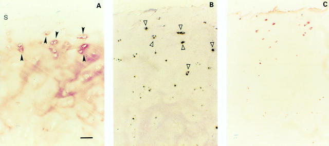
Figure 3 .
(A) Moderate OA cartilage (one of seven samples, Mankin 7). TSP-1 protein in moderate OA cartilage is present in the upper regions with a pericellular distribution, whereas in the deeper regions a slight interterritorial staining persists (s indicates cartilage surface). (B) Strong TSP-1 expression of the pericellularly stained chondrocytes was found by in situ hybridisation. (C) CD36 positive chondrocytes in co-localisation with TSP-1 expressing chondrocytes. Bar represents 125 µm in figs A, B, and C.

