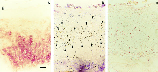
Figure 5 .
(A) Osteophytes. Strong immunohistochemical TSP-1 staining in osteophytes (s indicates cartilage surface). (B) Paralleled by a strong TSP-1 expression by in situ hybridisation (area between closed arrowheads). (C) Most of the chondrocytes are CD36 positive. Bar represents 125 µm in figs A, B, and C.

