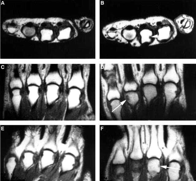
Figure 3 .
Magnetic resonance images of the three new erosions (white arrows) developed within the observation period of one year in three patients. T1 weighted, spin echo, pre-contrast axial images (A) at baseline, and (B) at the one year follow up; a new erosion is seen in the 3rd metacarpophalangeal (MCP) joint. (C) T1 weighted, spin echo, pre-contrast coronal image at baseline and (D) at the one year follow up, a new erosion is seen in the 4th MCP joint. (E) T1 weighted, spin echo, pre-contrast coronal image at baseline and (F) at the one year follow up, a new erosion is seen in the 3rd MCP joint.

