
Figure 1 .
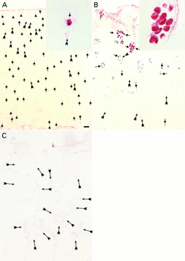
(A) Normal articular cartilage immunostained with antibodies against VEGF, showing positive (arrowheads) and negative chondrocytes (arrows) in all cartilage layers; Mankin score 1. VEGF positive and negative chondrocytes from the deep zone are shown in close up (inset). (B) Severe OA cartilage with a strong immunostaining of the chondrocyte clusters next to the surface (arrowheads) and also VEGF positive chondrocytes throughout the remaining deep zone (arrows); Mankin score 11. Positive chondrocytes within a cluster are displayed in close up (inset). (C) A slide adjacent to (B) incubated with non-immune rabbit serum as negative control. Arrows indicate negative chondrocytes. Bar represents: (A) 200 µm; (B) 100 µm; and (C) 60 µm.