
Figure 1 .
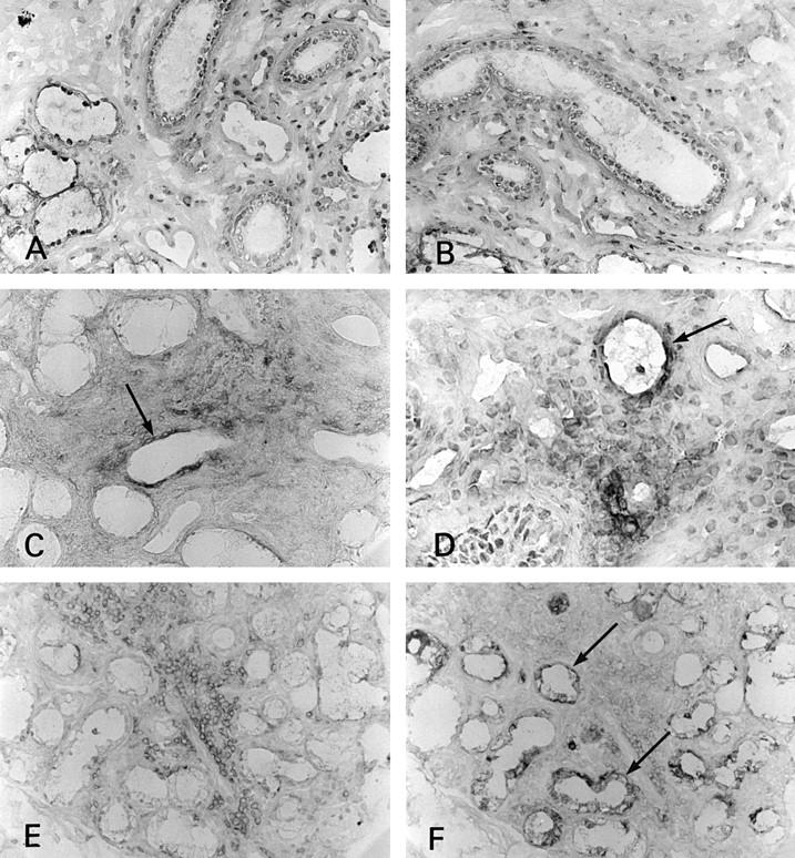
Immunohistochemical staining of CD80 and CD86 in the salivary gland. (A) CD80 was not seen in normal controls (×180); (B) CD86 was not seen in normal controls (×180); (C) CD80 on ductal epithelial cells (arrow) and some infiltrating mononuclear cells in severe sialoadenitis (×180); (D) CD86 was expressed on ductal epithelial cells (arrow) and some infiltrating mononuclear cells in severe sialoadenitis (×360); (E) CD28 was seen on many infiltrating mononuclear cells in severe sialoadenitis around the duct expressing CD80; (F) serial section of (E) (×180).