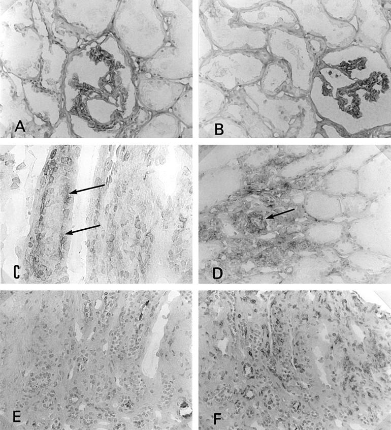
Figure 3 .

Immunostaining of CD80 and CD86 in the kidney. (A) CD80 was not seen on tubular epithelial cells in control tissue (×180); (B) CD86 was not seen on tubular epithelial cells in control tissue (×180); (C) CD86 expression on tubular epithelial cells (basal side, arrows) in interstitial nephritis associated with Sjögren's syndrome (SS) (×360); (D) the several tubular cells showed CD86 antigen in SS kidney (arrow) (×180); (E) CD80 was not seen on tubular epithelial cells in interstitial nephritis associated with SS (×180); (F) CD28 expressed on some infiltrating cells (the same portion of the serial section of (C) and (E) (×180)).
