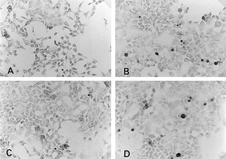
Figure 4 .

Immunohistochemical staining of anti-CD80 or CD86 antibody on cultured human salivary gland duct (HSG) cell line (×180). Without cytokine stimulation, CD80 (A) or CD86 (B) expression was not seen on HSG. Many HSG cells stimulated by 100 U/ml of interferon γ for 24 hours showed CD80 (C) or CD86 (D) expression.
