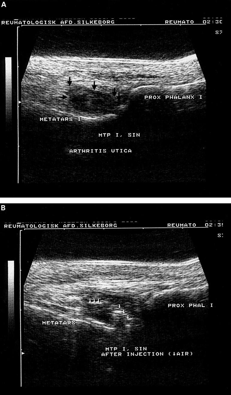
Figure 1 .

(A) Ultrasonography of a metatarsophalangeal (MTP) joint before injection. The distended capsule is seen (black arrows). (B) Ultrasonography of MTP joint after injection of sterile air (white arrows) and 2 ml steroid. Only the volume of air that is in the needle itself is injected (~0.05 ml).
