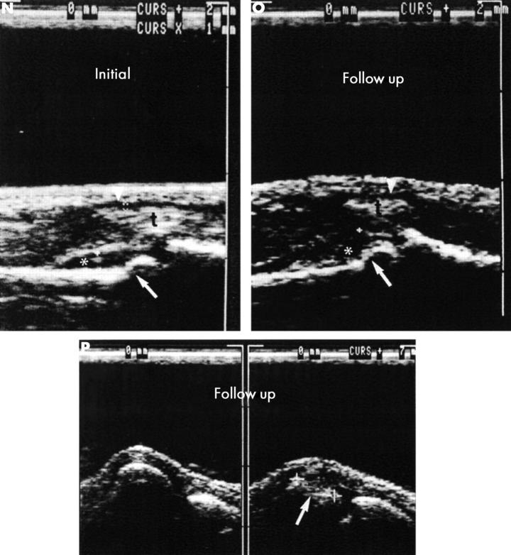
Figure 5 .
Images obtained by radiography (A-D), MRI (E-I), scintigraphy (J-M), ultrasound (N-P) of the clinically most severely affected left hand in a woman with RA aged 20 at the time of the initial examination (time 0) and 22 at the time of follow up. (A, B) Survey radiograph of the left hand at time 0 in dorsovolar projection (A) and with the hand in the volardorsal, semisupine (45°) position with abducted fingers, the so-called "zither player position" (B): demonstration of a narrowed joint cleft in PIP joint III and of a small cystoid brightening on the radial side without disruption of the border lamella at the head of the proximal phalanx of digit III (open arrow). There is partial loss of the subchondral border lamella at the ulnar base of the proximal phalanx of digit IV (closed arrow). These changes do not yet represent direct signs of arthritis (Larsen stage 1). (C, D) Follow up radiography of the left hand performed two years after the initial examination in the dorsovolar projection (C) and in the zither player position (D): the joint clefts of all PIP joints now clearly show narrowing. The cystoid brightening at the head of the proximal phalanx of digit III seen at time 0 is now definitely identified as an erosion, but only on the film obtained in the zither player position (thick arrow) and not on the dorsovolar projection. The partial loss of the subchondral border lamella at the ulnar base of the proximal phalanx of digit IV now shows recalcification. Newly developed cystoid lesions not disrupting the border lamellae (thin arrows) are seen on the radial side of the head of the proximal phalanx of digit II, on the ulnar side of the head of the proximal phalanx of digit V, at the metacarpal head of digit I, and in corresponding locations at the scaphotrapezoid joint. The new erosion in PIP joint III is a direct sign of arthritis affecting less than 25% of the joint area, corresponding to Larsen stage 2.

