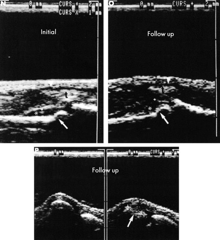
Figure 5 .
(contd) (E, F, G) MRI of the left hand performed at time 0 using an unenhanced T1 weighted 3D gradient echo sequence. The figure only shows one coronal section (E) obtained before contrast administration and a corresponding postcontrast section (F, G). (E) A large erosion on the radial side of the head of the proximal phalanx of digit III (open arrow) can be seen, which is demonstrated by MRI in association with extensive synovitis (asterisk) at PIP III already two years before there is clear cut radiographic evidence of its presence (fig 5D). After contrast medium administration (F), this erosion is partly obscured by the pronounced enhancement of the synovitis (*) at PIP III. Additional, smaller erosions—likewise not demonstrated radiographically—are seen in the metacarpal head of digit IV (arrow with loop) (more clearly seen in other images), on the ulnar side of the head of the proximal phalanx of PIP IV (thin, white arrow), and on the ulnar side of the metacarpal head of digit V (white arrow ) (G). Florid, contrast-enhanced synovitis (*, s) is depicted at PIP joints I–V, and at MCP joints I, II, IV, V. Florid, contrast-enhancing tenosynovitis (t) is seen affecting the flexor tendons of fingers 1–5. The radiographically demonstrated (fig 5A) partial loss of the subchondral border lamella at the ulnar base of the proximal phalanx of digit IV corresponds to contrast-enhancing osteitis affecting the entire base of the phalanx at MRI (black arrow). (H, I) After two years of DMARD treatment the patient underwent follow up MRI of the left hand using the same parameters as at time 0 using a comparable section orientation. The known lesion on the radial side of PIP joint III shows regression but still shows pronounced enhancement reflecting florid activity. The erosion on the ulnar side of PIP joint IV (open arrow (I) shows progression in the presence of more extensive synovitis (*, I). The tenosynovitis on the flexor side of digit III has a higher signal intensity on the postcontrast image than at time 0.

