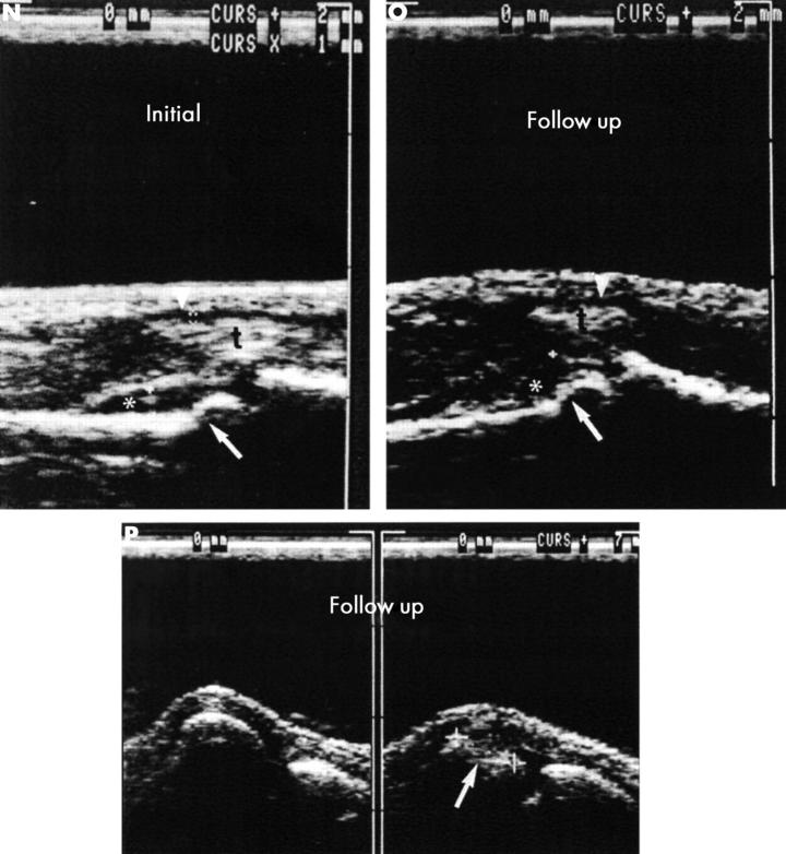
Figure 5 .
(contd) (J, K) Initial scintigraphy, phases II (J) and III (K), shows hot spots in PIP joints II, III, and IV and in the wrist (lunate bone or adjacent parts of ulnar and radial bones). (L, M) At follow up two years later, scintigraphy, phase II (L), shows hot spots in the wrist, MCP joints I and III, IP joint, PIP joints II and III. Scintigraphy, phase III (M), shows hot spots in the wrist, MCP joints I, III, and V, interphalangeal joint, and DIP joints II, III, and IV.

