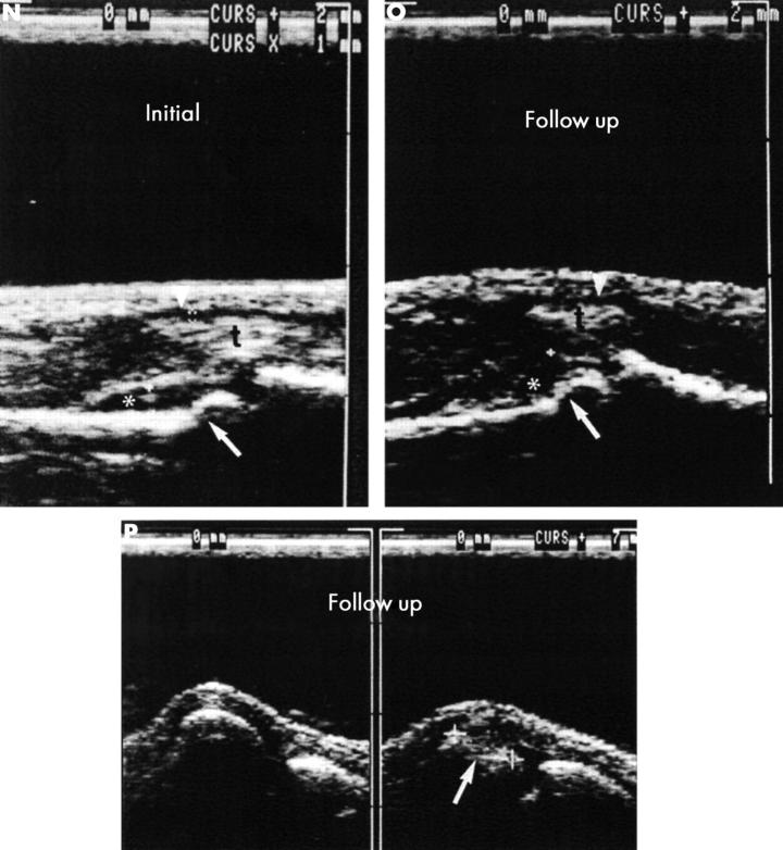
Figure 5 .
(contd) (N, O, P) (N) The initial ultrasound examination showed synovitis of the proximal interphalangeal (PIP) joints II, III. Ultrasound shows a hypointense line indicating synovitis (*) in PIP joints II (left) and III (right). The flexor tendon sheaths (arrowheads) of the second and third fingers are distended as an indication of tenosynovitis (t, flexor tendon). The step-like interruption of the bone margin (arrow) of the head of the proximal phalanx of digits II and III is indicative of erosion. (O) The figure shows a hypointense line indicating synovitis (*) in PIP joints II (left) and III (right). The flexor tendon sheaths (arrowheads) of the second and third fingers show minimal distention as an indication of tenosynovitis (t, flexor tendon). Irregularities of the joint contour were found at the heads of the proximal phalanx II and III (arrows). Ultrasound shows a large erosion at the head (arrow) of MCP joint V (P, right side) in the dorsal aspect and in the flexed MCP joint, which was not seen at conventional radiography (figs 5A-D) but in both MRI examinations (initially and at the two year follow up, fig 5G). The left side shows a normal joint contour of MCP joint IV.

