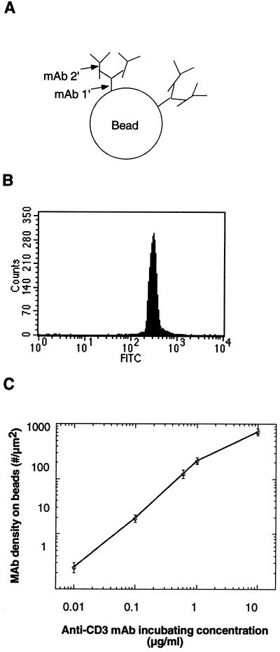
Figure 2.
Quantification of immobilized FITC-conjugated anti-CD3 mAb density on polystyrene beads by FACS analysis. (A) Antibody coating on beads. The two-step antibody coating method was assumed to prevent potential steric hindrance and increase anti-CD3 mAb binding efficacy. Beads were initially coated with 100 μg/ml anti-hamster Fc IgG mAb (mAb 1′) and then with varying concentrations of FITC-conjugated hamster anti-mouse CD3 mAb (mAb 2′) of known fluorescein/protein ratio. mAb 1′ binds the Fc portion of mAb 2′, leaving the antigen-binding site of mAb 2′ free to engage with the TCR:CD3 complex. (B) FACS analysis of beads shown in A. The number of anti-CD3 mAb on single beads was quantified by using FACS analysis to compare FITC-conjugated mAb-coated beads with a standard curve of microbeads labeled with defined numbers of fluorescein molecules. (C) The resulting anti-CD3 mAb density on beads showing a 3-log linearity with incubating antibody concentration.

