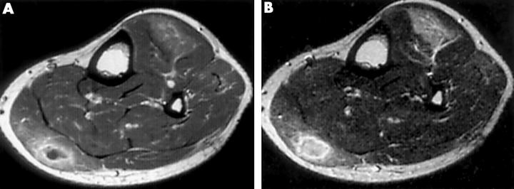
Figure 1 .
MRI of the lower legs was performed on the patient's first admission. An axial T1 weighted image showed a focal mass-like lesion, about 3 cm in diameter, in a left gastrocnemius muscle with decreased intensity relative to that of normal muscle. After administration of gadolinium, the T1 weighted image showed a well defined rim of contrast enhancement and a hypointense central area (A). An axial T2 weighted image showed bright signal intensity in and around a focal mass-like lesion (B).

