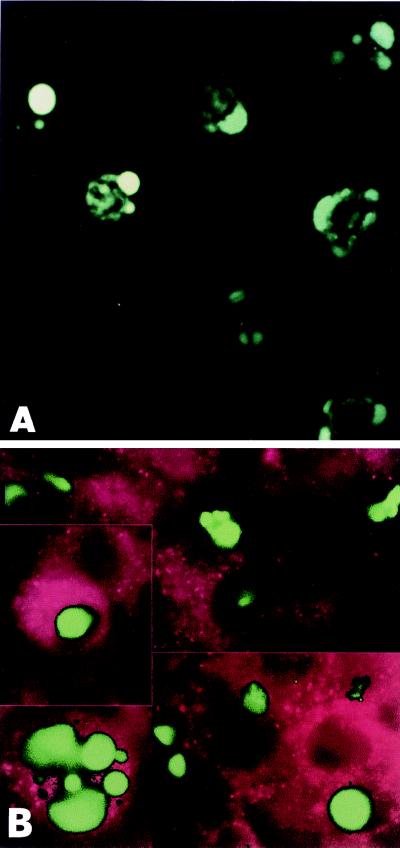
Figure 1.
(A) In situ TUNEL staining of clone 2.I. Condensed and fragmented nuclei incorporated the fluorescein-labeled dUTP, thus indicating apoptosis-induced DNA strand breaks. (B) Double-immunofluorescent labeling of Fhit-reexpressing cells. Apoptotic nuclei are FITC-labeled. Fhit cytoplasmic expression is detected by rhodamine-conjugated polyclonal anti-Fhit antibody.

