
Figure 3.
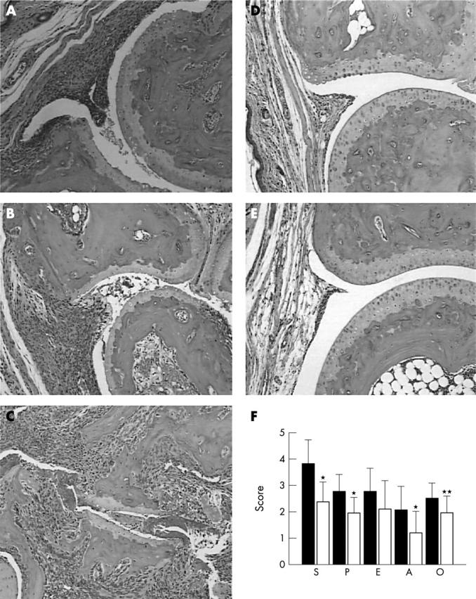
Histopathology of arthritis in B6.IFNγ KO mice. (A) Section of a hind paw with arthritis at the day of onset showing inflamed synovium with massive infiltrate of mononuclear cells. There is decreased number of chondrocytes in the articular cartilage. (B) Section of a hind paw with arthritis at day 14 showing severe synovitis, pannus formation, and erosion and some visible destruction of articular cartilage. (C) Section of a hind paw with arthritis at day 35 showing massive synovitis, erosion of cartilage and subchondral bone, and destruction of joint structure. (D) Normal appearance of a section of a hind paw from a non-arthritic CII immunised B6 mouse. (E) Section of a remitted arthritic hind paw from a B6 mouse showing normal joint appearance. (F) Histological assessment of arthritis in affected limbs in B6.IFNγ KO mice (open bars) at the end of the experiments (day 35 of arthritis) in comparison with that in DBA/1 mice (filled bars). S, synovitis; P, pannus formation; E, erosion; A, architectural changes; O, overall histological score. *p<0.01; **p<0.05.